
Physio Performance, Health & Fitness
What Causes a Frozen Shoulder? The Physio Guide to Understanding Adhesive Capsulitis
Frozen shoulder causes are one of the more frustrating topics in musculoskeletal medicine, because in a large proportion of cases there is no identifiable trigger at all. A patient who has done nothing unusual, injured nothing and changed nothing develops progressively worsening shoulder pain and stiffness over several months.
That said, the underlying process is reasonably well understood, and the risk factors are clear enough to be genuinely useful. This guide explains what actually happens inside the joint, who is most at risk, what triggers it where a trigger exists, and what the honest limits of current understanding are.
What Happens Inside the Joint
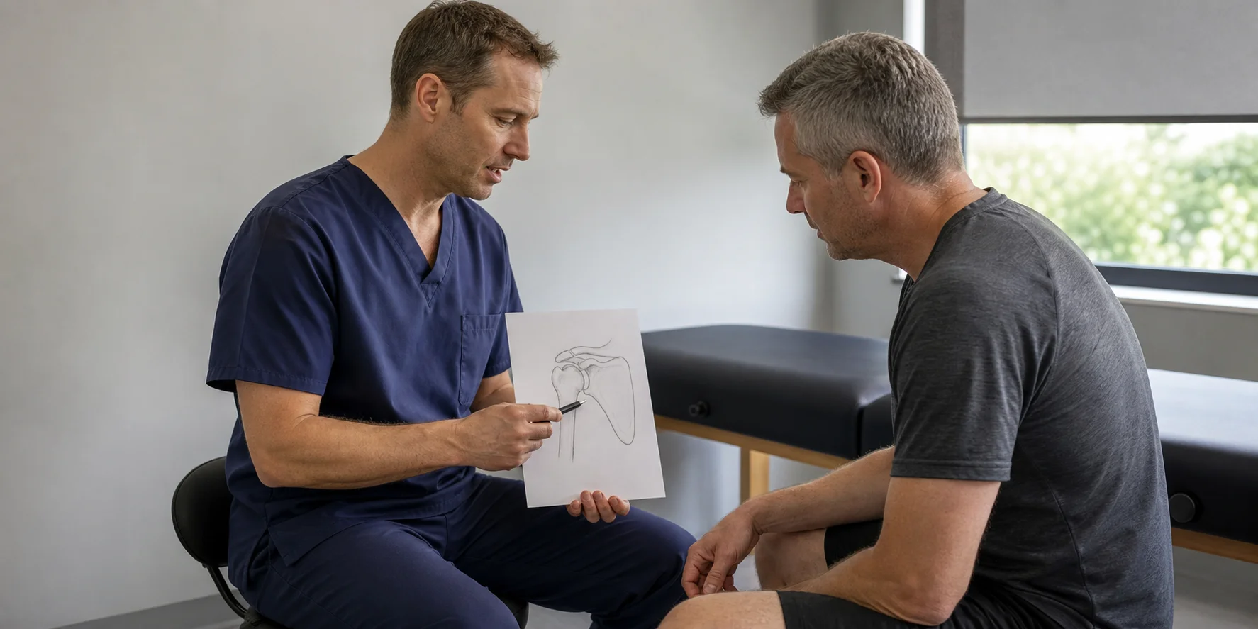
The shoulder joint is surrounded by a capsule, a sleeve of connective tissue that holds the joint together while allowing the very large range of movement the shoulder requires.
In adhesive capsulitis, that capsule undergoes two changes. First it becomes inflamed, which produces the pain that dominates the early stage. Then it thickens and contracts, forming fibrous tissue that physically restricts movement. The capsule effectively shrinks around the joint.
This explains the characteristic pattern. Because the capsule surrounds the joint entirely, restriction occurs in all directions rather than in specific movements. And because the restriction is structural rather than muscular, it does not matter whether you move the arm or someone else does. The block is the same.
It also explains why external rotation is typically the worst affected. The portion of the capsule most involved in the contracture, at the front of the joint, is precisely the part that must lengthen for the arm to rotate outward.
The Established Risk Factors
Diabetes. By some distance the strongest association. People with diabetes have a substantially elevated incidence, and cases tend to be more severe, more resistant to treatment and longer lasting. Bilateral involvement is also more common. The mechanism is thought to relate to how elevated blood glucose affects collagen structure, though this is not fully settled.
Age between forty and sixty. The condition is strongly age-banded. Presentation outside this range, particularly in someone considerably younger, warrants closer investigation for other causes.
Thyroid disease. Both overactive and underactive thyroid are associated with increased incidence.
Period of immobility. Any circumstance that keeps the arm still for several weeks, whether a fracture, surgery, or a soft tissue injury treated with prolonged rest, raises risk considerably. This is one of the reasons modern practice avoids prolonged shoulder immobilisation wherever possible.
Previous frozen shoulder on the other side. A meaningful proportion of people who have had it once develop it in the opposite shoulder, usually within five years.
Cardiovascular disease and stroke. Both are associated, likely partly through reduced arm use.
Parkinson’s disease. Also associated, again likely related to reduced movement.
Our guide on frozen shoulder symptoms covers how to recognise the condition once it develops.

What Triggers It Where There Is a Trigger
Secondary frozen shoulder is the term for cases with an identifiable precipitating event, and roughly speaking these fall into a few categories.
Following a shoulder injury. A rotator cuff strain, a fall onto the shoulder, or any injury that caused a period of reduced movement. Notably the initial injury is often relatively minor. It is the reduced movement afterwards rather than the injury itself that appears to matter.
Following surgery. Shoulder surgery obviously, but also breast surgery, cardiac surgery, and any procedure that limits arm use during recovery.
Following a fracture. Particularly wrist fractures, where the arm is immobilised in a cast for several weeks. Frozen shoulder after a Colles fracture is common enough to be a recognised complication.
Following a period of illness. Any illness involving prolonged bed rest or reduced arm use.
The pattern across all of these is reduced movement over a period of weeks. This is why current practice emphasises early mobilisation after shoulder and arm injuries wherever it is safe, and why prolonged sling use is avoided unless genuinely necessary.

Primary Frozen Shoulder: The Honest Answer
In a substantial proportion of cases there is no identifiable trigger whatsoever. This is termed primary or idiopathic frozen shoulder.
Patients frequently find this the most difficult part to accept. There is a natural assumption that a significant problem must have a cause, and the absence of one feels like something has been missed.
What is understood is that an inflammatory process begins in the capsule and progresses to fibrosis. What is not fully understood is what initiates that process in someone who has done nothing to provoke it. Research points toward inflammatory and fibrotic pathways with possible genetic contributions, but there is no clear single answer at present.
The practical implication is that in primary cases there is usually nothing you did to cause it and nothing you could have done to prevent it. That is worth saying clearly because patients frequently spend considerable energy searching for a cause that does not exist.
Clinical guidance on shoulder conditions is published by the National Institute for Health and Care Excellence, and current research on the pathology appears in the British Journal of Sports Medicine.
Can Frozen Shoulder Be Prevented
For primary frozen shoulder with no identifiable trigger, there is no established prevention strategy. This is a straightforward answer that patients deserve rather than vague reassurance.
For secondary frozen shoulder following injury or immobility, the picture is more useful. Maintaining shoulder movement during any period where the arm is otherwise restricted meaningfully reduces risk.
Practically that means gentle pendulum movements and assisted range work during recovery from a wrist fracture, after shoulder or chest surgery where the surgical team permits it, during any illness involving reduced activity, and after any arm injury requiring a sling.
The instruction is not to load the shoulder but simply to move it. A few minutes of gentle movement several times daily is sufficient and substantially better than complete immobility.
For anyone with diabetes, glycaemic control appears relevant given the association, though the evidence that improving control reduces frozen shoulder risk specifically is not conclusive.
Why Understanding the Cause Matters for Treatment
Knowing the underlying process directly shapes the correct approach.
Because the early stage is inflammatory, aggressive stretching during that phase increases irritation and prolongs the condition. Because the later stage is fibrotic contracture, sustained gentle stretching in that phase does produce genuine change. This is why matching treatment to stage is so important, as covered in our guide on exercises for frozen shoulder.
Because the restriction is capsular rather than muscular, strengthening exercises do not restore range and mobility work is required. Strengthening becomes relevant later once range returns.
And because the process is self-limiting but slow, the goal of treatment is shortening the course and preserving function rather than curing something that would otherwise be permanent. Our guide on how long frozen shoulder lasts covers the timelines.
If you have developed shoulder pain and stiffness and want an accurate diagnosis and stage assessment, book an appointment at our clinic in Drogheda. You can also download our free neck and shoulder pain report for interim guidance.
Patient information on shoulder conditions is published by the Chartered Society of Physiotherapy.
Frequently Asked Questions
The capsule surrounding the shoulder joint becomes inflamed and then thickens and contracts, physically restricting movement. In many cases there is no identifiable trigger. Where there is one, it is usually a period of reduced arm movement following injury, surgery or illness.
The association is strong and well established, thought to relate to how elevated blood glucose affects collagen structure in connective tissue. Cases in people with diabetes also tend to be more severe and longer lasting.
There is no established causal link between psychological stress and adhesive capsulitis. Stress can increase muscle tension and pain perception, but it is not recognised as a cause of the capsular changes that define the condition.
Indirectly. A relatively minor injury that results in several weeks of reduced arm movement can precipitate it. It appears to be the immobility rather than the injury itself that matters, which is why early gentle movement after arm injuries is encouraged.
Research suggests possible genetic contributions to susceptibility, and some families do show clustering. However it is not considered a straightforwardly hereditary condition and most cases occur without family history.
Related reading
Struggling with this? See our shoulder pain treatment in Drogheda.